Third ion imaging workshop 2022 in Munich, Germany - Summary

Summary of event attendance
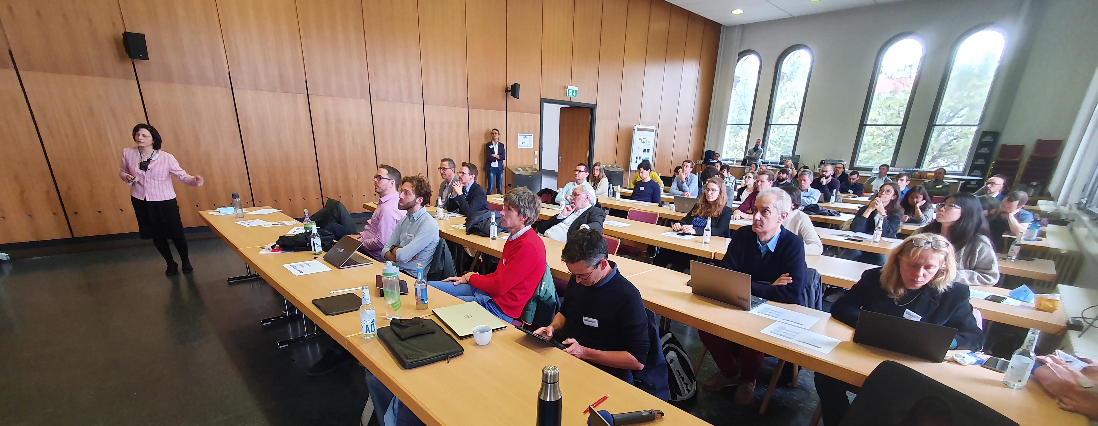
The 3rd Ion Imaging Workshop took place on October 13th and 14th at the LMU main building in Munich and was following workshops in Lyon in 2018 and Manchester in 2019. The workshop attracted 50 registered participants, which marked an increased participation compared to Lyon (36 participants) and Manchester (42 participants). The workshop had an international character, with participants from Norway, France, Italy, Germany, Switzerland, the United Kingdom, the Netherlands, Austria, and the United States. External funding was instrumental in ensuring a well-attended workshop, allowing the organizing committee to invite speakers.
In total, 26 presentations were given in the 8 sessions below:
- Ion imaging systems 1
- Small animal systems
- Reconstruction methods
- Combined and novel modalities
- Performance of ion imaging systems and clinical application
- Ion imaging systems 2
- Radiography
- Theoretical investigations and modelling
The event was endorsed by ESTRO, and the journal Physics in Medicine and Biology has opened a focus collection on the topic of ion imaging where workshop contributions can be published as full papers.
Scientific summary of the event
On October 13th, the event opened with the first session on ion imaging systems, where invited talks by Prof. Dieter Röhrich of Norway and Prof. Albert Hirtl of Austria presented the development of their respective list-most proton computed tomography scanner designs. The audience learned about the operation the Norwegian digital calorimeter based on disentangling hits, aiming at a high count rate which could yield a proton computed tomography (pCT) system fast enough to be clinically applicable. Prof. Hirtl elaborated on their concept to use the time of flight of protons as a data acquisition method for pCT. Dr. Carlo Civinini from INFN Florence presented the latest results from their pCT prototype scanner, confirming that this system can achieve clinically relevant image quality goals.
The second session of the morning covered the recent developments of small animal systems, where invited speaker Prof. Katia Parodi presented the SIRMIO project. The audience also learned from Dr. Palaniappan (presented by Prof. Marco Riboldi due to illness) about 2D-3D image registration of small animals using proton radiography, which indicates a way for incorporating proton radiographies (pRad) into the clinical workflow of proton therapy.
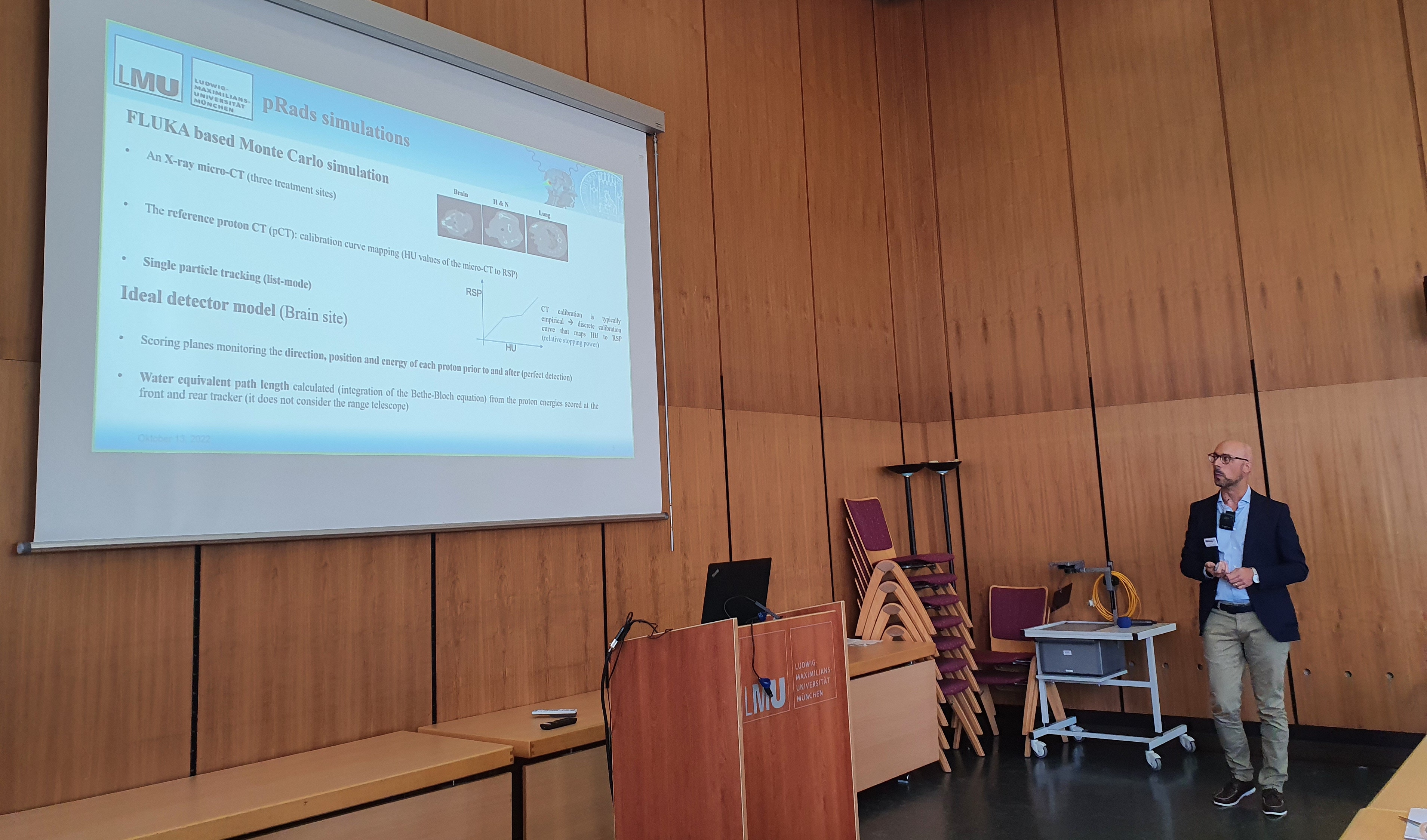
After lunch presentations on image reconstructions showed recent developments. Dr. Rit presented a new methodology for increasing spatial resolution in pCT images. Dr. Simard showed his latest results on how to acquire pRads from a monolithic detection system by reading out lateral 2D projections. Dr. Volz demonstrated in his talk how the well-established distance driven binning method for pCT could be adapted to pRad, allowing to optimally increasing resolution in proton radiographs acquired with single particle tracking systems. Closing that session, Ms. Götz presented a comprehensive study on the comparison of proton vs. He CT, based on various image quality metrics.
The final session of the day visited combined and novel imaging modalities, where invited speaker Dr. Chiara Gianoli presented the use of AI in ion imaging reconstruction. Invited speaker Prof. Joao Seco broadened the horizon with considerations of imaging secondary particles for FLASH radiotherapy monitoring. Finally, invited speaker Prof. Bruzzi presented recent work on how to improve the characterization of patient tissue required for proton therapy, using pCT.
On October 14th the day opened with a session on clinical applications of ion imaging, with an invited talk from Prof. Antje Knopf on clinical use of proton radiography with an integrating detector. During that talk, Prof. Knopf shared with the audience some of the first human patient radiographies, indicating the potential of this technique for being translated to a clinical methodology. Ms. Fogazzi further elaborated on the concept presented the previous day by Prof. Bruzzi, highlighting how current state of the art pCT scanners could improve patient tissue characterization required for proton treatment planning. The last talk of the session, given by Ms. Seller Oria, explained how pRad is an attractive technique for benchmarking AI based improvements of the current clinical workflow. In general, the session explored how ion imaging could have a near-term impact in clinical proton therapy.
The second session of the morning focussed on the second set of presentations on ion imaging systems. Invited speaker Prof. Robert Johnson presented a comprehensive overview of all existing and functional proton imaging systems, highlighting the current challenges with the development of next generation ion imaging systems. Invited speaker Prof. Sam Beddar, an expert on clinical dosimetry with plastic scintillators, discussed how this detector technology can be exploited for ion imaging. Finally, Dr. Ulrich-Pur elaborated on recent detector developments enabling the design and construction of a time of flight based pCT system.
After lunch a session on radiography saw novel approaches from Los Alamos National Laboratory with a talk from Dr Martin Schanz on the use of magnetic lenses for ion imaging and discussed the challenge of how to adapt a very large imaging system designed for different purposes to a smaller, clinically applicable pCT scanner. The audience also heard about imaging with helium ions from the DKFZ team with talks from Yanting Xu, focusing on how this can be used for patient alignment, and Margareta Metzer, presenting a quantitative analysis of the image quality of helium radiography. Mr. Ryan Fullarton closed that session by explaining in detail the detector design and performance of a monolithic silicon detector system for pRad.
In the final session of the meeting, Ms. Stefanie Kaser presented an overview of the ion imaging research performed at the Austrian particle therapy facility. Dr. Nils Krah, presented a detailed theoretical investigation of the novel concept of time-of-flight ion. Dr George Dedes presented a performance comparison of two ion imaging prototypes, which were based on different design concepts.
The two-day meeting closed with the remarks of the organizing committee and announced the next ion imaging workshop, to be held in 2023 in London. In the discussion, which took place during that last part, a few new inter-institutional projects were, identified. Of particular interest was the idea to organize and execute a cross prototype comparison based on well-characterized phantoms, since several well performing ion imaging systems are now available.